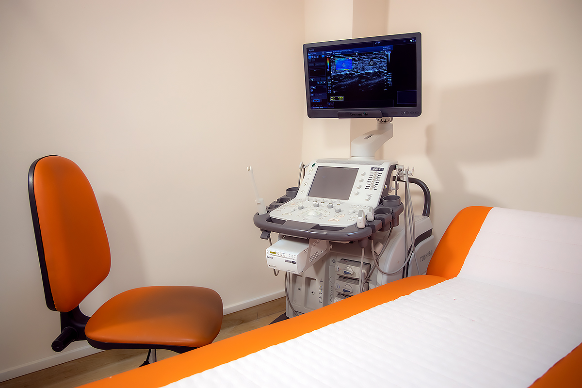
Ultrasound diagnostics 3D & 4D
Equipment for ultrasound diagnostics in Slavmed Medical Center is a part of the latest achievements in modern medicine because it is equipped with 3D / 4D ultrasound system, which is one of the best technologies of the new generation visualization. The system of special options ensures sharpness and clarity of tissue image. The device is equipped with new Fly Thru and Inversia special options that can be compared with virtual endoscopy as it enables the physician to see the slots of hollow organs, ducts and arteries as well as to implement the visualization of the mentioned organs from outer area, which is called Inversia. By this method it is possible to receive additional information on tumors and invasive growth of dimensional formations. Ultrasound Examination (US) provides both morphological and functional characteristics of organs and organ systems by providing qualitative and quantitative data. The device enables you to detect very small tumors, accurately examine their internal structure, get clearer images and description of minor tumors and surrounding tissues, thus increasing the efficiency of diagnostics and distinctive diagnostics. This is a very good opportunity to discover diseases in early stage. Due to pain and risk-free nature of the examination, it can be performed with no time and quantity limitation thus allowing full control of the course and development of the disease.
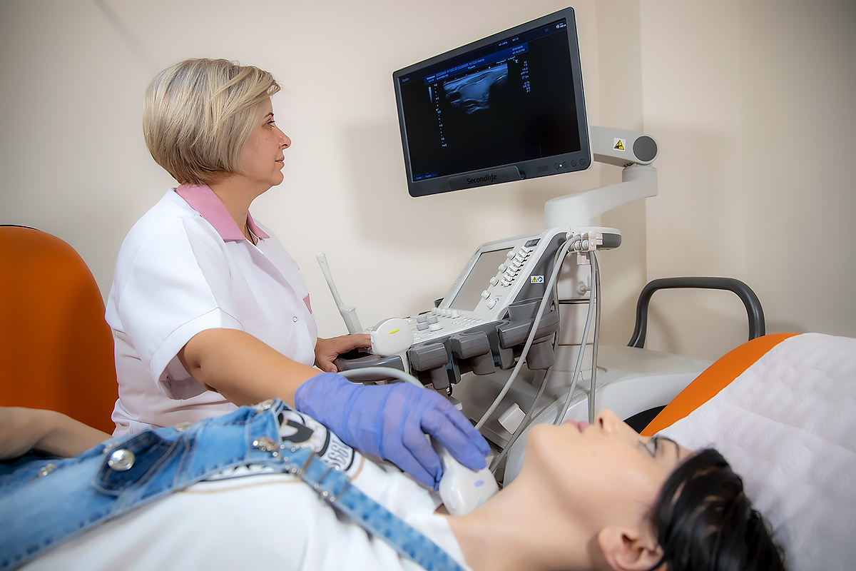
The highly qualified specialists in the field of radiology of Slavmed Medical Center are regularly trained at leading foreign medical institutions and present their reports during major conferences and seminars.
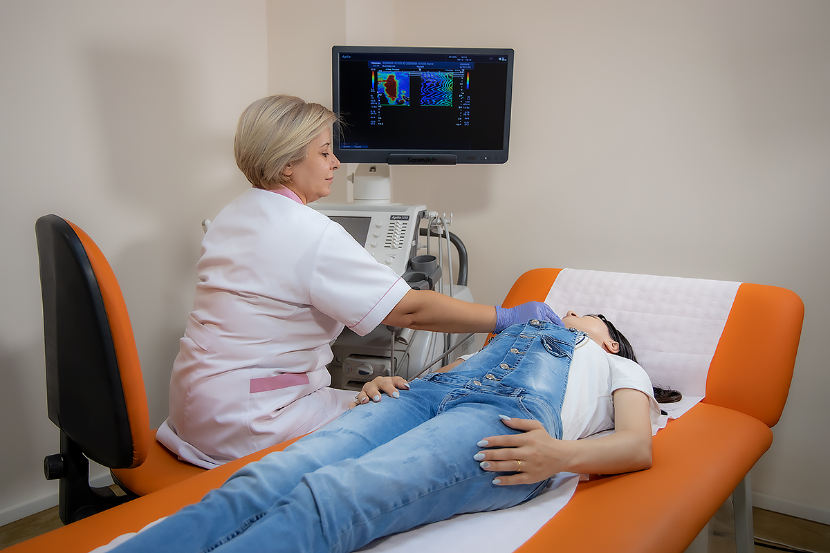
Ultrasound examination of the abdomen
It is a diagnostic ultrasound examination of the abdominal organs: gallbladder, liver, spleen and pancreas. It allows not only to examine in detail the functional and anatomical characteristics of the organs and systems of the patient’s abdomen, but also to find cause-and-effect relationship of present abnormalities, thus enabling to properly describe the condition of organs being examined, based on the patient complaints and the clinical picture.
Thanks to abdominal ultrasound it is possible to confirm accurate sizing of all internal organs, describe their structure, discover or rule out the presence of any site of inflammation and tumors.
An abdominal ultrasound in performed in the following cases:
– gallstone disease,
– liver cirrhosis,
– hepatitis,
– chronic or acute cholecystitis,
– chronic or acute pancreatitis,
– tumors (benign or malignant),
– abdominal aortic aneurysm
Acute pain in abdomen is also an indicator that US of the abdominal area is needed.
This type of examination can also be used to estimate the effectiveness of an undergoing treatment. The accuracy of the ultrasound examination of the abdominal cavity is very important because the gases in the intestine can deteriorate the visibility of the organs that are being examined, which can affect the results of the examination. Doctors of Slavmed Medical Center recommend getting an abdominal ultrasound done early in the morning, on an empty stomach. In case it has to be performed in the afternoon hours, it is recommended to have a light breakfast in the morning, as long as there is a six-hour break between breakfast and ultrasound.
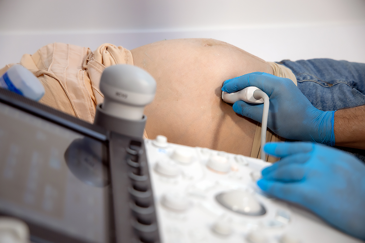
Ultrasound in Obstetrics & Gynecology
Ultrasound examination allows to confirm pregnancy on the earliest stages, research the anatomy of fetus, its overall condition, to perform prenatal diagnostics, folliculometry, overall vessel Doppler velocimetry of womb, placenta-fetus as well as diagnose diseases of womb and ovary, research pelvic organs.
Renal ultrasound
The examination allows discovery of fluid-containing mass in kidney, kidney tumors, hyponephrosis, urolithiasis, presence of tumors and concrements and Doppler examination allows to evaluate renal vessels condition. An ultrasound examination helps to discover not only stones bladder and urethral lumen stones, but also ptosis of ureters and bladder tumors. Ultrasound examination of testis, scrotum and prostatic gland allows to diagnose varicocele diseases as well as to determine the degree of blood flow in case of a twisted testicle. Ultrasound examination is invaluable to confirm a testicular cancer diagnosis in order to make it sure there is a blood circulation in the injured scrotum. An ultrasound of prostatic gland allows discovering the affected area, determining the size of prostate, which provides important information regarding early detection of benign prostatic hyperplasia (adenoma) and cancer. Ultrasound helps to discover misperfusion of penis and to determine the degree of blood flow.
For additional information please contact our administrators directly via phone number or e-mail address provided below:
Phone: +374 (10) 322211 (open 24/7)
info@slavmed.am